Research
Projects
Investigations across cell biology, genome architecture, and neuroimmunology. Click any project to expand.

Biological underpinnings of the Matthew effect
Tracing how early-life adversity compounds into lifelong immune and neurodevelopmental vulnerability.
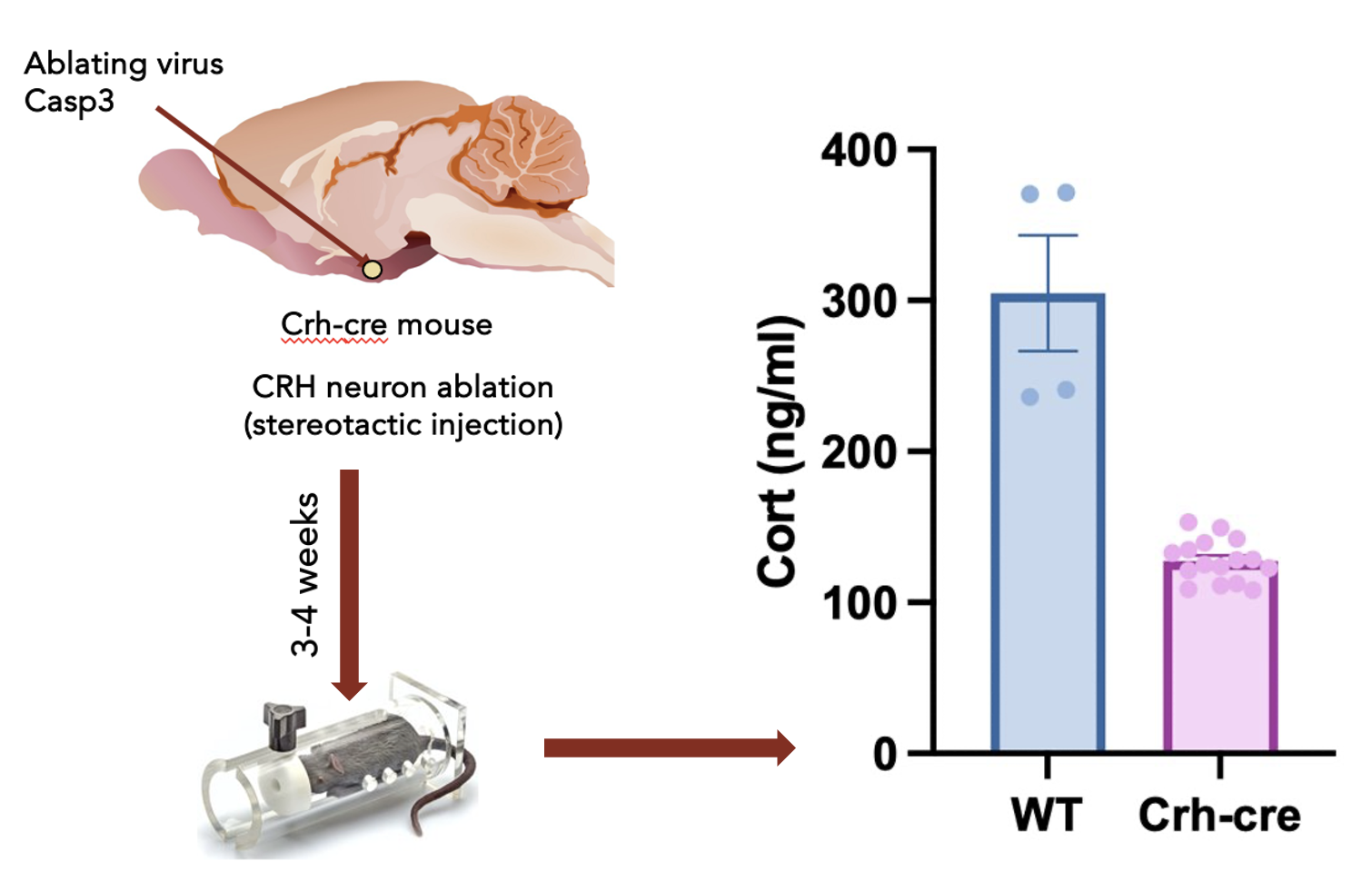
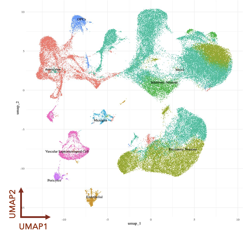
The Matthew Effect describes how early disadvantages (or privileges) compound over a lifetime. Children born to stressed mothers face elevated disease risk that does not reset at birth and can be transmitted transgenerationally, as documented in the children conceived or born during the Dutch Hunger Winter. Understanding the biological mechanisms that embed these early-life adversities into lasting disease risk is both a scientific and a public health imperative. Using single-cell transcriptomics, multiplexed proteomics, and neurologic interventions in mice, I investigate how maternal stress reshapes immune function and brain development in offspring, and how these changes propagate across generations. The goal is to identify points of intervention that can reduce, if not reverse, the long-term health consequences of prenatal stress. This work is part of the Biology of Adversity Project at the Broad Institute. Figures : Top: Matthew effect: The rich get richer; poor, poorer. Middle: Stereotaxic ablation of stress centers in the mouse brain, confirmed by reduced cortisol levels. Bottom: Single-cell transcriptomic clustering of mouse brain cells.
Seligowski, Dhuppar, et al., (in preparation)
miRNAs in Cancers
Characterizing the roles of miR-92a and miR-21 in colorectal cancer, colitis, and Multiple Sclerosis.
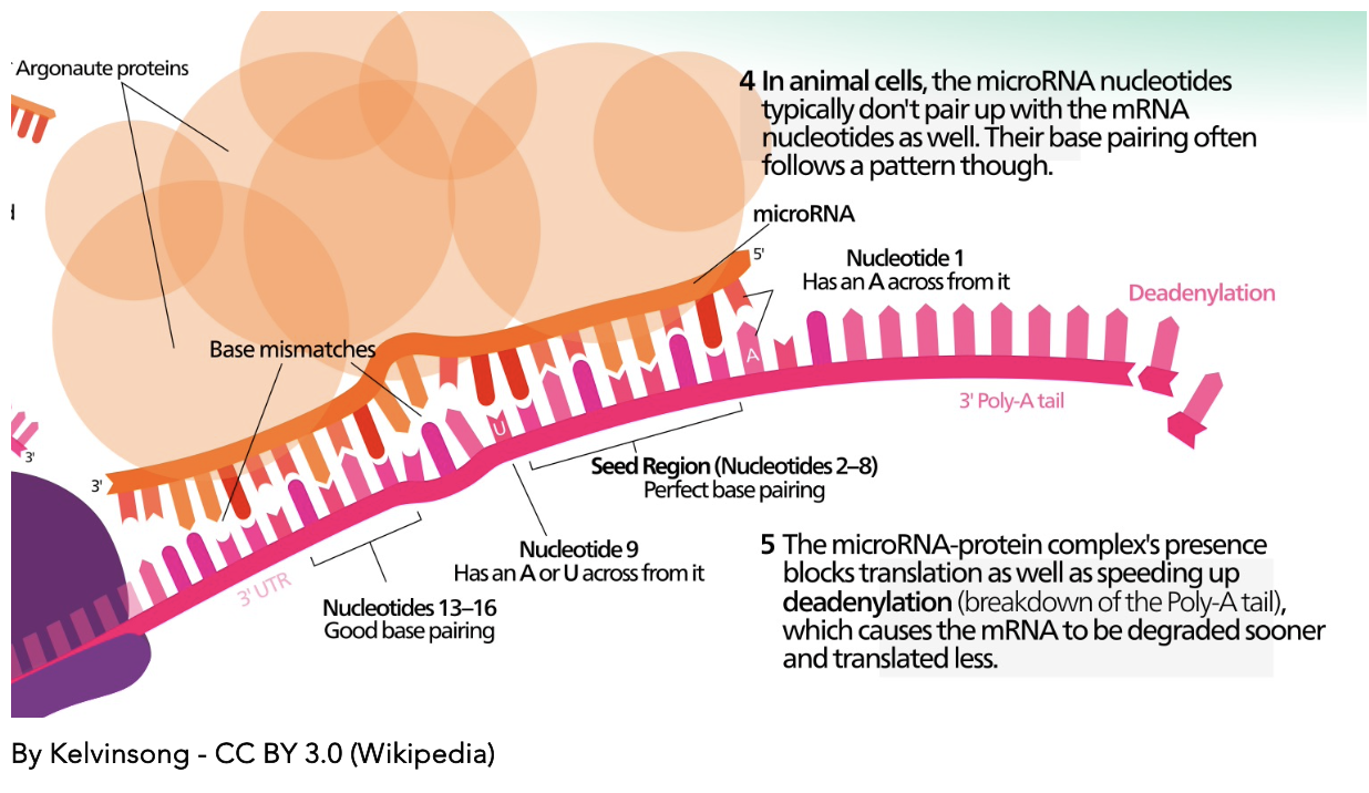
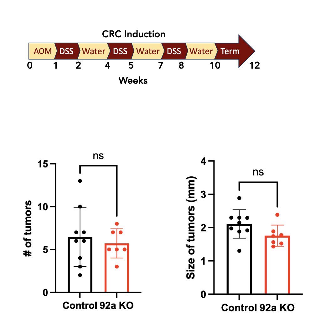
MicroRNAs are small non-coding RNAs that regulate gene expression post-transcriptionally. Their discovery earned Gary Ruvkun and Victor Ambros the 2024 Nobel Prize in Physiology or Medicine. Ruvkun, at this institution, has spent decades unraveling how these molecules control development and disease. miR-92a, from the oncogenic miR-17-92 cluster, promotes colorectal tumor growth by suppressing pro-apoptotic and anti-proliferative targets, and modulates inflammatory signaling in the gut. Using knockout mouse models and high-dimensional immune profiling of the lamina propria, I investigated how its loss reshapes immune composition during colitis. In a separate collaborative project, we show that Type I interferon limits CNS autoimmunity through the miR-21–FOXO1 axis in pathogenic Th17 cells, providing a mechanistic basis for IFN-β therapy in Multiple Sclerosis. Figures: Top: RISC-mediated mRNA degradation and translational repression by miRNAs. Bottom: Reduced tumor susceptibility in miR-92a KO mice
Varghese et al. Science Translational Medicine, 2025 Dhuppar, Poller, and Gopal, Trends in molecular Medicine, 2025 Dhuppar and Gopal, Trends in Immunology, 2022
Cell Cycle Staging from Microscopy
Automated image analysis pipeline to stage cells in the cell cycle without chemical synchronization.
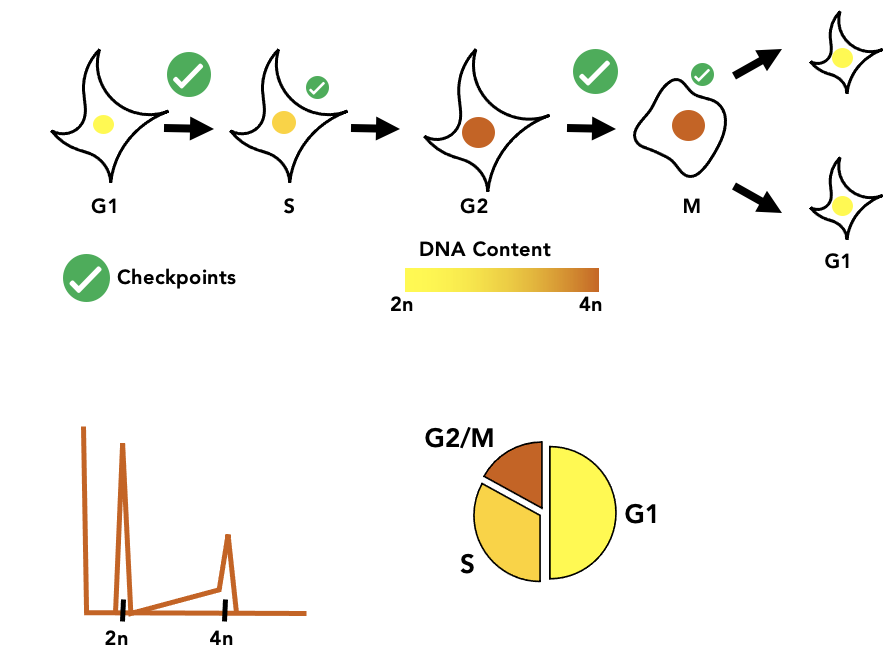
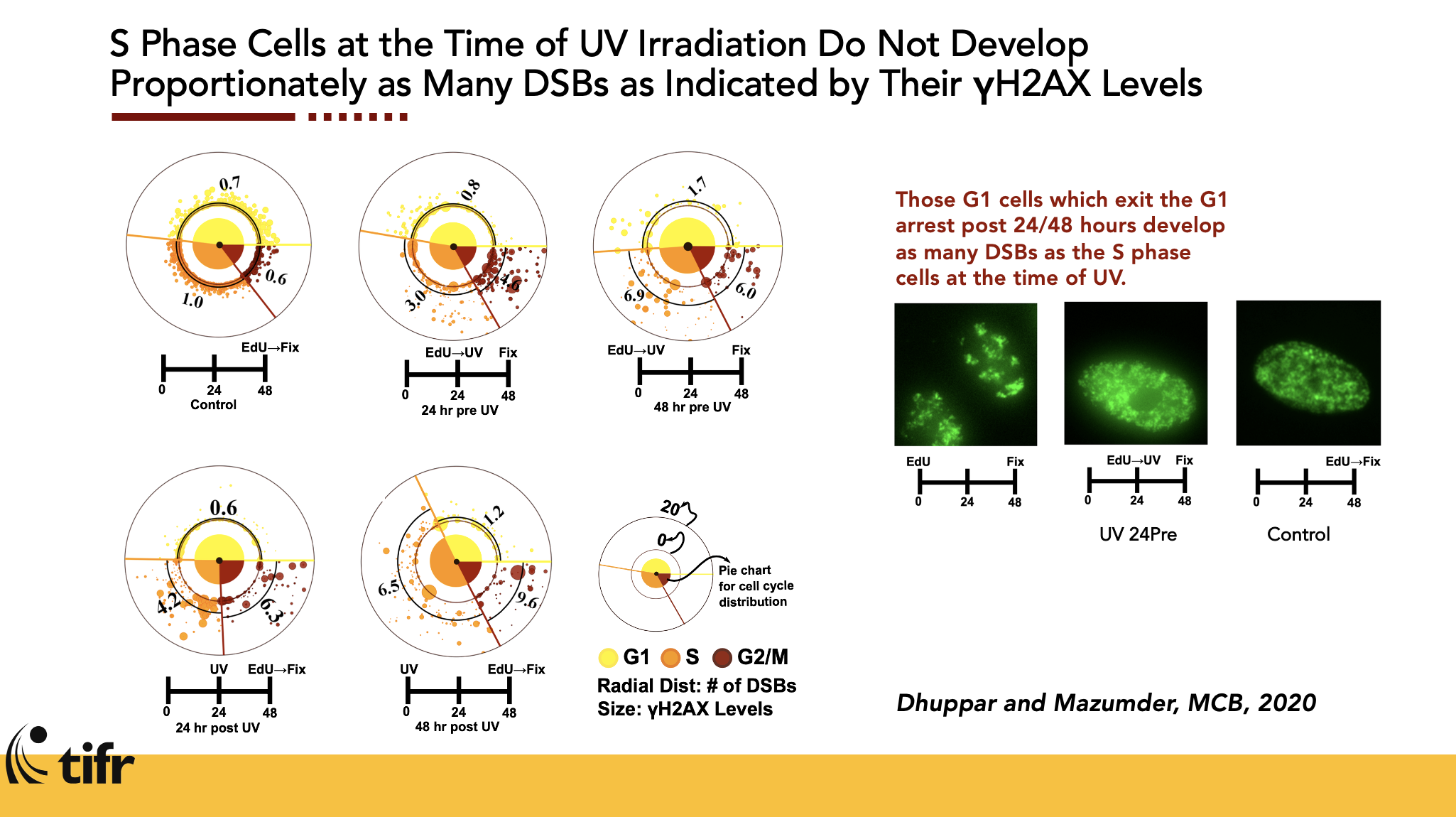
Chemical synchronization of cells, the standard approach to cell cycle studies, perturbs the very biology under investigation. I developed a fully automated image analysis pipeline to stage individual cells in the cell cycle from high-resolution fluorescence microscopy images, eliminating this confound entirely. Combining DNA FISH, RNA FISH, and immunofluorescence, the pipeline enabled single-allele-resolution studies of gene expression and DNA damage responses. One key finding: the γH2AX peak in S phase after UV irradiation marks sites of active DNA replication, not the extent of damage itself, with direct implications for how DNA repair is interpreted in cycling cells. Figures: Top: Image-based cell cycle staging against DNA content by flow cytometry. Bottom: Cell Cycle-dependent response to UV-induced DNA damage.
Dhuppar, Roy & Mazumder. Mol Cell Biol, 2020 Dhuppar & Mazumder. Cell Cycle, 2018 Dhuppar & Mazumder. J Cell Science, 2020
Genome Architecture & Gene Regulation
How CTCF-mediated three-dimensional genome organization governs monoallelic gene expression.
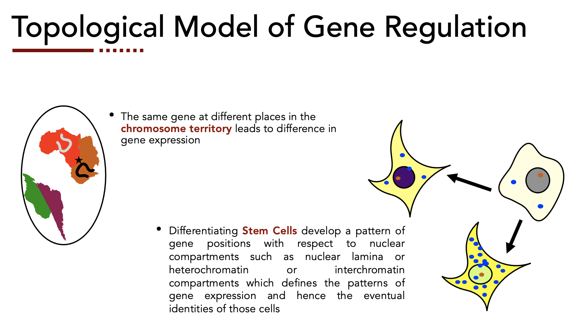
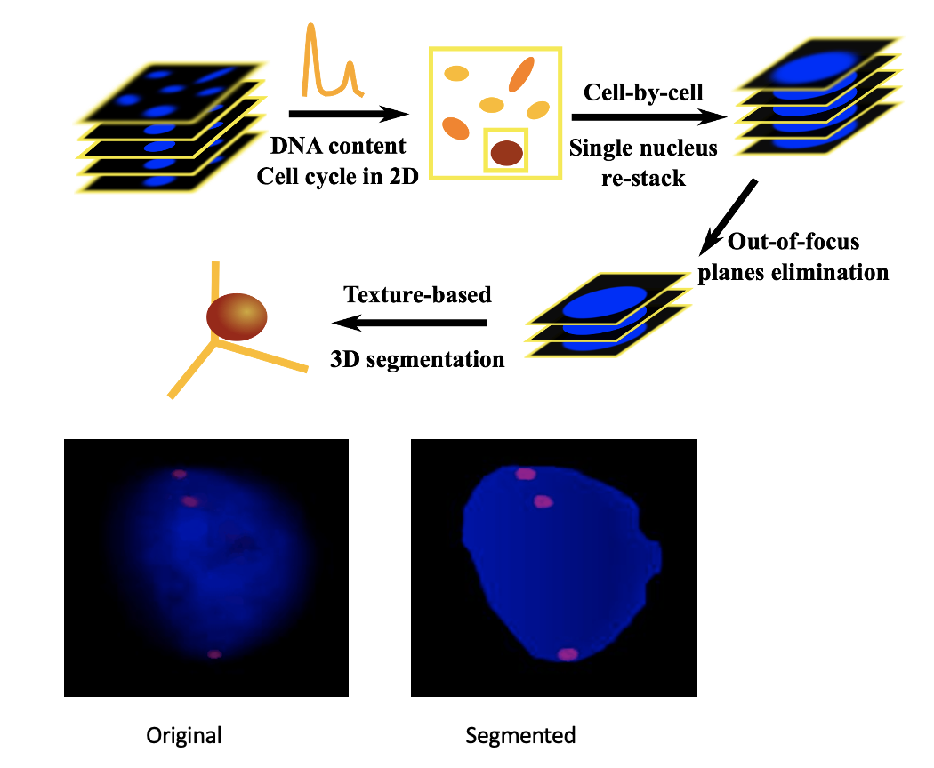
As cells differentiate, genes reposition relative to nuclear compartments, including the lamina, heterochromatin, and interchromatin regions, in patterns that define cell identity and regulate expression. Whether these positional relationships hold during cell cycle progression, when chromatin undergoes large-scale reorganization, was less clear. Using DNA and RNA FISH at single-allele resolution, I showed that key genes are transcribed independently of their position relative to the nuclear lamina across cell cycle phases, challenging a simple repression model. In related collaborative work, CTCF-mediated genome architecture was shown to govern dosage of monoallelic autosomal gene expression. I extended these themes at the Children's Hospital of Philadelphia, using Hi-C to map cell cycle-dependent changes in three-dimensional chromosome organization. Figures: Top: Topological model of gene regulation. Bottom: Completely-automated, high-throughput image analysis pipeline for studying nuclear architecture-based gene regulation at single-allele resoltion.
Sen, Dhuppar, & Mazuzmder, FISH Methods and Protocols, 2024 Dhuppar & Mazumder. J Cell Science, 2020 Chandradoss, Chawla, Dhuppar et al. Cell Reports, 2020
Publications
* marks corresponding authorship
Full Publications List
2018 – present
click to expand